Testing & Diagnosis
What a qEEG can and can't tell you after a brain injury
The short answer
qEEG measures brain electrical patterns. It supports, but doesn't replace, a neurologist's exam and history. That doesn't mean it can reliably diagnose a concussion. Current guidance doesn't support qEEG for confirming or excluding mild TBI. Any proposed test should answer a specific question and be interpreted with its limitations clearly explained.
What is qEEG actually measuring?
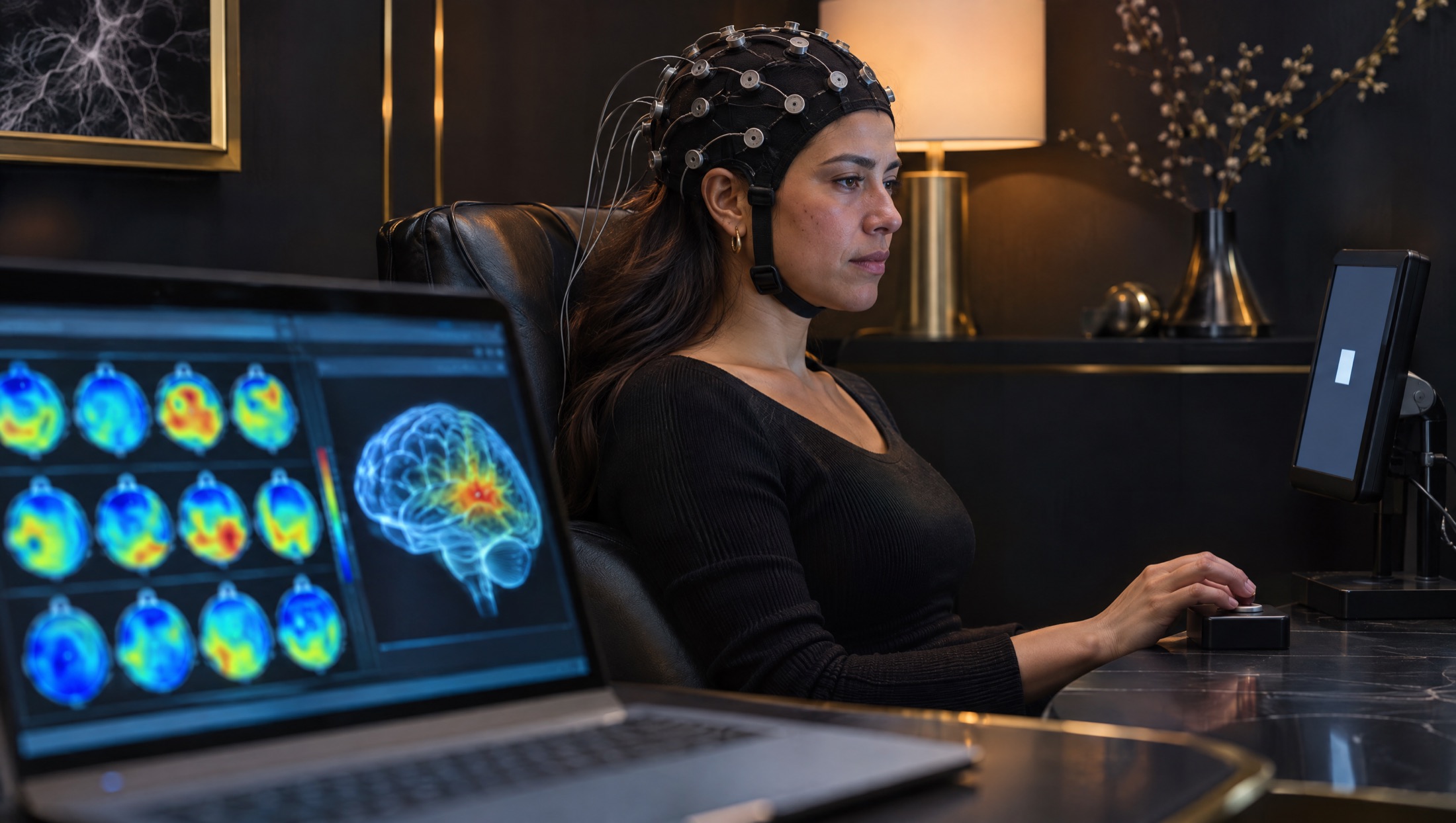
An electroencephalogram, or EEG, records electrical activity through electrodes placed on the scalp. Quantitative EEG applies mathematical analysis to that recording. It can summarize features such as the amount of activity in different frequency ranges. Some reports display those summaries as colored maps or compare them with a reference group.
Those displays are measurements and calculations, not photographs of damaged brain tissue. A red area isn't a bruise, and a blue area isn't a hole. Before interpreting a map, the clinician needs to understand how the signal was recorded and processed. The National Library of Medicine's EEG overview explains the basic recording process and why the patient's condition during the test matters.
Is qEEG accurate for brain injury diagnosis?
That question needs a narrower definition of accurate. A system can measure an electrical feature consistently without being able to identify what caused it. For patients asking, “Is qEEG accurate for brain injury?”, the practical issue is whether the result can reliably distinguish injury from other explanations and improve a clinical decision.
The American Clinical Neurophysiology Society's practice guideline found that the evidence did not support qEEG to diagnose mild TBI, either at the time of injury or later. It also found insufficient evidence to distinguish mild TBI from other diagnoses or to identify it in people taking central nervous system medications. The guideline characterizes this diagnostic use as investigational.
That conclusion matters when a test is marketed as proof that an accident injured the brain. A report can contain numbers and still leave the central clinical question unanswered. Ask the ordering clinician to explain the evidence for the specific use being proposed, rather than relying on a broad claim about brain mapping.
What can a careful assessment of electrical activity contribute?
Conventional EEG has established uses, including assessment of suspected seizures. In some clinical settings, quantitative displays can help trained clinicians examine large amounts of EEG data. That use is different from applying a statistical brain map to diagnose persistent symptoms after a mild head injury.
If you've had unexplained spells, episodes of unresponsiveness or possible seizures after a crash, your neurologist may recommend an EEG for that specific concern. Ask whether the test is looking for seizure activity, summarizing the recording or making a claim about concussion. Those are separate questions and may require different evidence.
A useful discussion starts with what would happen after either result. If a finding would prompt another examination or a change in management, ask why. If the plan would remain exactly the same, ask what additional value the test provides. You're entitled to understand the purpose before deciding whether to proceed.
What can't a qEEG report establish on its own?
A qEEG map can't independently establish that a particular crash caused a particular symptom. It can't determine an exact injury date or turn a statistical difference into a precise measure of disability. It also can't promise how quickly you'll recover or identify a treatment that is certain to work.
A normal result doesn't rule out a concussion or make persistent symptoms unimportant. An abnormal result doesn't automatically identify traumatic damage. Either result needs to be considered alongside the event, your medical history, examination findings and other clinically relevant information. A test shouldn't replace listening to the patient.
Nor is a map a substitute for emergency assessment. New weakness, repeated vomiting, a worsening headache, a seizure or difficulty waking after head trauma needs emergency care. The CDC's danger sign guidance applies whether a previous test looked normal or abnormal. Don't wait for a scheduled brain mapping appointment when those symptoms appear.
Why can recording conditions change the result?
An EEG records small electrical signals at the scalp, where other activity can interfere. Eye movements, blinking, muscle tension, movement and electrode contact can affect a recording. Being awake, drowsy or asleep also changes normal brain rhythms. Medicines and other health conditions can influence the signal.
The person interpreting the study should evaluate the underlying EEG and recording quality, not just the finished graphic. Ask how artifacts were identified and handled. If a reference database is used, ask whether it is appropriate for the person being tested and the recording method. A comparison is only meaningful when its assumptions are reasonable.
Follow the laboratory's preparation instructions. Bring a complete medication list and tell the team about poor sleep or other circumstances that might matter. Don't stop medication or deliberately deprive yourself of sleep unless the ordering clinician instructs you to. Preparation should support safe testing, not produce a preferred result.
Does FDA clearance mean a device can diagnose every concussion?
No. FDA status applies to a particular device and its intended use. It isn't a blanket endorsement of every EEG method, analysis service or marketing statement. The FDA's TBI guidance warns against using devices outside appropriate authorization for head injury assessment and explains that diagnosis involves clinical information.
Ask for the device name and what its authorization actually covers. Does it apply to your age, the time since injury and the decision being considered? A tool intended to help assess an acute injury isn't automatically validated for explaining symptoms months later. The clinician should be able to describe that distinction without relying on promotional language.
How does qEEG fit with scans and neurological testing?
Different tools answer different questions. CT can help detect acute bleeding when clinically indicated. MRI provides anatomical information. EEG records electrical activity. A neurological examination assesses functions such as strength, sensation, coordination and eye movements. None of these is interchangeable with the others.
The CDC's clinical guidance for mild TBI provides a framework for assessment and follow-up. Persistent symptoms may call for evaluation of headache, sleep, balance, vision, mood or cognitive difficulties. The selection of tests should follow those clinical questions rather than a fixed package offered to everyone after a crash.
Bring earlier reports so the clinician can avoid repeating work without a reason. Ask what information is still missing. If you receive conflicting interpretations, request a written explanation of the uncertainty or another specialist opinion. You shouldn't have to choose between believing your symptoms and asking reasonable questions about a test.
What questions should Texas patients ask before scheduling?
Start with the decision the clinician is trying to make. Then ask who will interpret the study, whether the raw EEG will be reviewed and whether the result is expected to change care. Ask about limitations as directly as benefits. A balanced explanation should cover both.
Discuss practical matters before the appointment: preparation, duration, cost, insurance coverage and how you'll receive the report. Ask whether there are other reasonable options, including clinical follow-up without the test. Don't assume an impressive report guarantees reimbursement or that a larger testing package means better care.
NeuroLUX's EEG service information describes a different clinical question from its qEEG information. Use those pages to prepare questions for your neurologist. For accompanying neck or back symptoms, SpineLUX explains musculoskeletal evaluation. Keeping those concerns visible can help prevent every symptom being attributed to one test result.
What else do patients ask?
Can qEEG prove that my headaches came from the crash?
Not by itself. The history, headache pattern, examination and other possible causes all matter. A statistical difference in an electrical recording doesn't establish the cause or date of an injury.
Does an abnormal map mean permanent brain damage?
No. A colored map isn't a direct image of tissue damage and doesn't independently predict permanence. Ask what the finding means, how reliable it is and whether it changes the clinical plan.
Can I still receive care if I don't have qEEG?
Yes. Evaluation and symptom management don't generally depend on having a qEEG. A clinician can assess your history and examination, address specific problems and recommend tests when they answer a useful question. Seek urgent care for new danger signs.
What should I do with a report I don't understand?
Ask for a plain language explanation and a copy to share with your treating clinician. Write down which conclusions are established, which remain uncertain and what the next step is. You don't need to interpret the color scale yourself.
Which sources inform this article?
General education only. This article cannot diagnose an injury or replace an examination. Care and recovery depend on individual clinical findings.
Written by Sung Won, MD. Published Nov. 4, 2026.